Dental X-ray
Types of X-rays we use
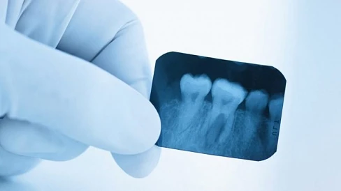
- Detects cavities between teeth, condition of fillings, roots, and bone
- Useful for treatment monitoring and follow-up
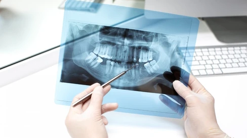
- Ideal for initial examination and treatment planning
- Useful before extractions, implants, and orthodontic treatment
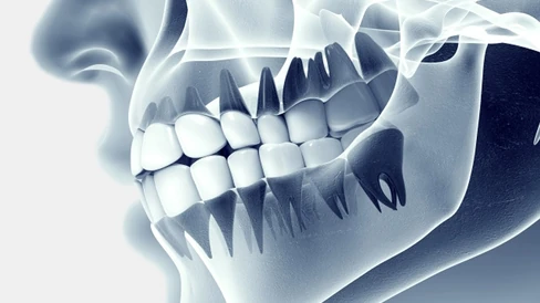
- Precise evaluation of bone, canals, and impacted teeth
- Helps identify risks and choose the safest treatment approach
When each type of X-ray is needed
Toothache / suspected cavity
Usually a periapical X-ray of 1–2 teeth is sufficient.
Treatment planning or initial examination
We often start with a panoramic X-ray (OPG).
Wisdom tooth or complex extraction
Depending on the case, an OPG or CT scan is used to assess roots and proximity to the nerve.
Implant placement / complex surgery
A CT scan is usually required for precise 3D planning.
Safety and radiation dose
Is it safe?
Modern dental X-rays involve very low radiation doses and extremely short exposure times.
We always choose the minimum required type of imaging for each clinical situation.
Pregnancy, childhood, and other individual factors are always discussed personally.
Important — If you have previous X-rays (OPG or CT scans), please bring them — often there is no need to repeat imaging.
Approximately “how much is it”
How the procedure works
Discussing the problem
We talk about your symptoms and clarify what needs to be assessed for diagnosis or treatment planning.
Taking the X-ray
You take a comfortable position while we fix the sensor or set the position — exposure lasts a fraction of a second.
Reviewing the results
We show the image on the screen and explain in clear terms what we see and what treatment options are available.
Treatment
After reviewing the image, we either proceed with treatment immediately or schedule it at a convenient time for you. You clearly understand what will be done and why.